58+ Asbestos x ray findings
Table of Contents
If you’re looking for asbestos x ray findings images information linked to the asbestos x ray findings keyword, you have come to the ideal site. Our website always provides you with hints for viewing the maximum quality video and picture content, please kindly surf and find more informative video articles and graphics that match your interests.
Asbestos X Ray Findings. Pleural thickening will usually be diagnosed based on the following findings. Tumors can also distort the normal shape of the lungs which can be detected on the radiograph. When a tumor is present on the pleura doctors will see a wispy white area that indicates tumor growth. Tap onoff image to showhide findings.
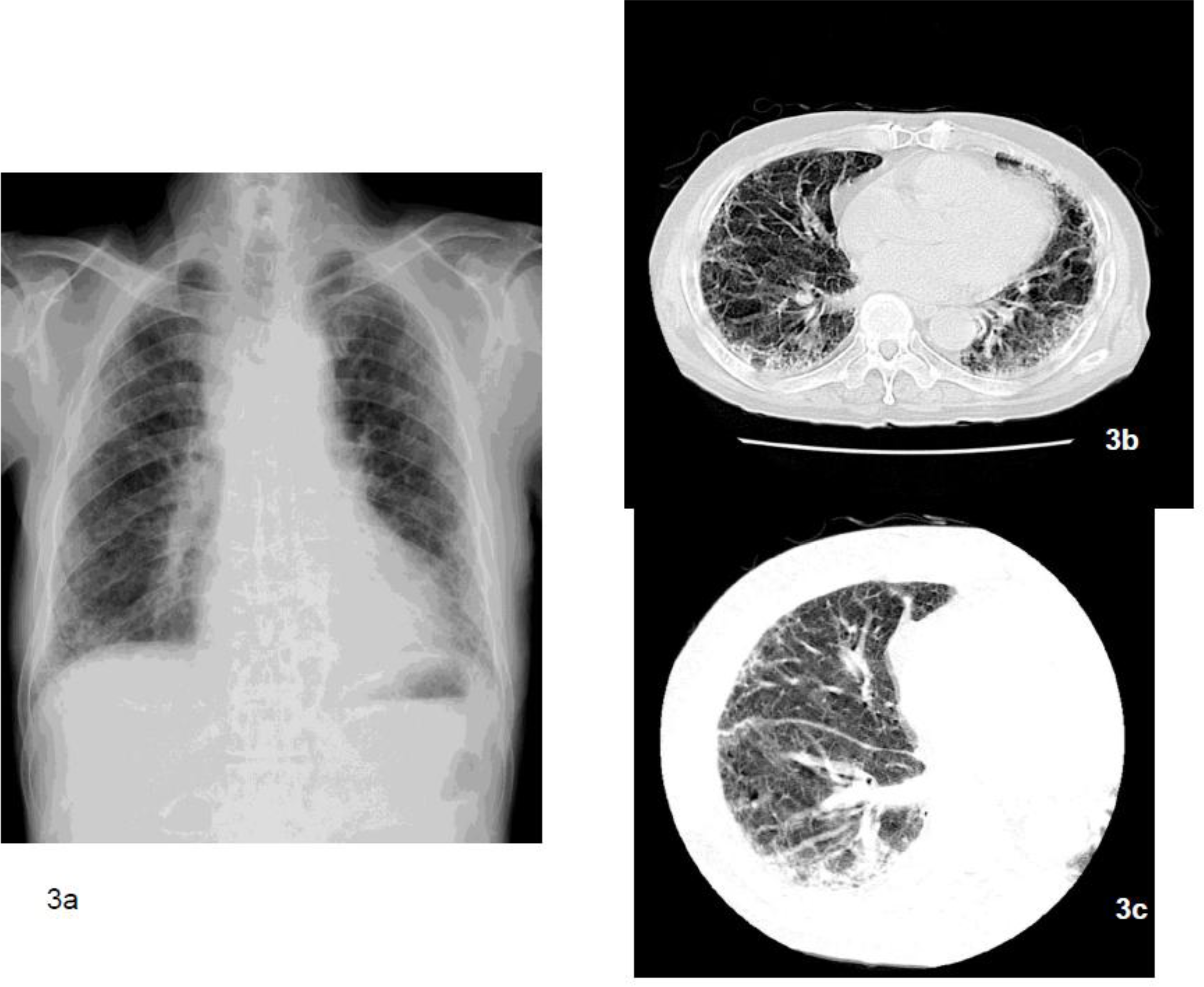 Ijerph Free Full Text Clinical Radiological And Pathological Investigation Of Asbestosis Html From mdpi.com
How many asbestos fibers cause cancer Asbestos xrd How many asbestos fibers are dangerous For asbestos abatement
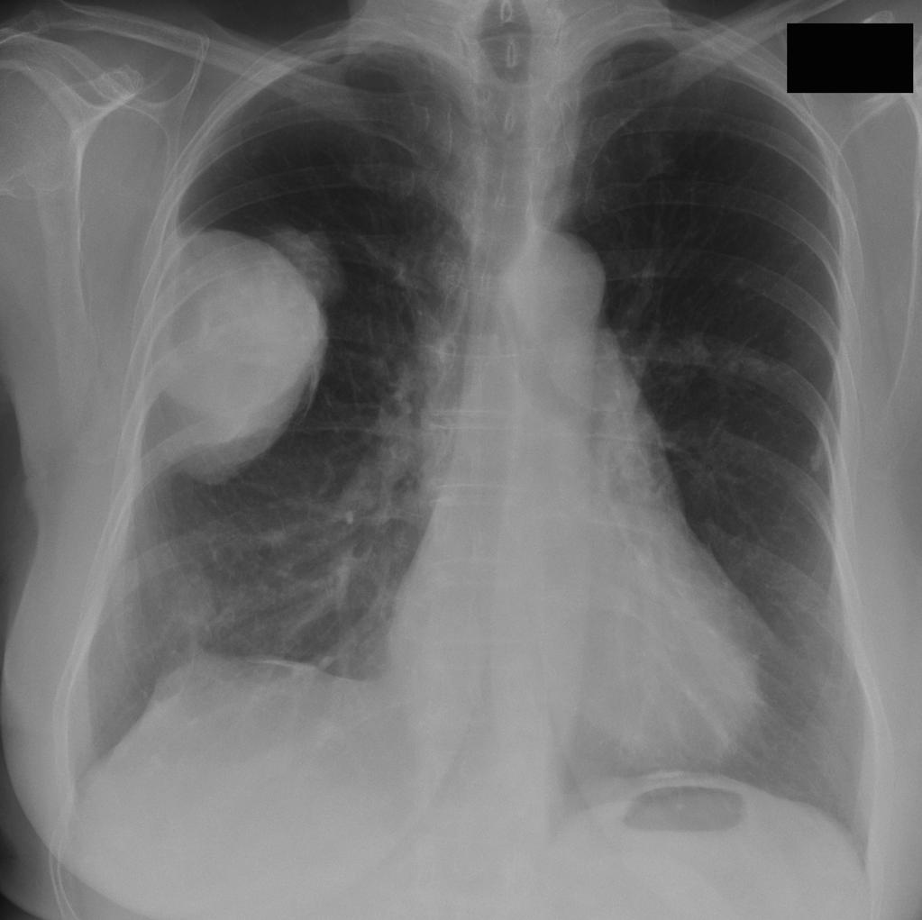
X Ray finding opaque masses along the lung walls. On standard X-rays healthy lungs appear black. Round atelectasis asbestos pseudotumor. This was confirmed by chest surgery. In both cases the symptoms can. The diaphragm is often the best place to look for plaques where they lie in the plane of the X-ray beam.
CT can help identify the disease in its early stages.
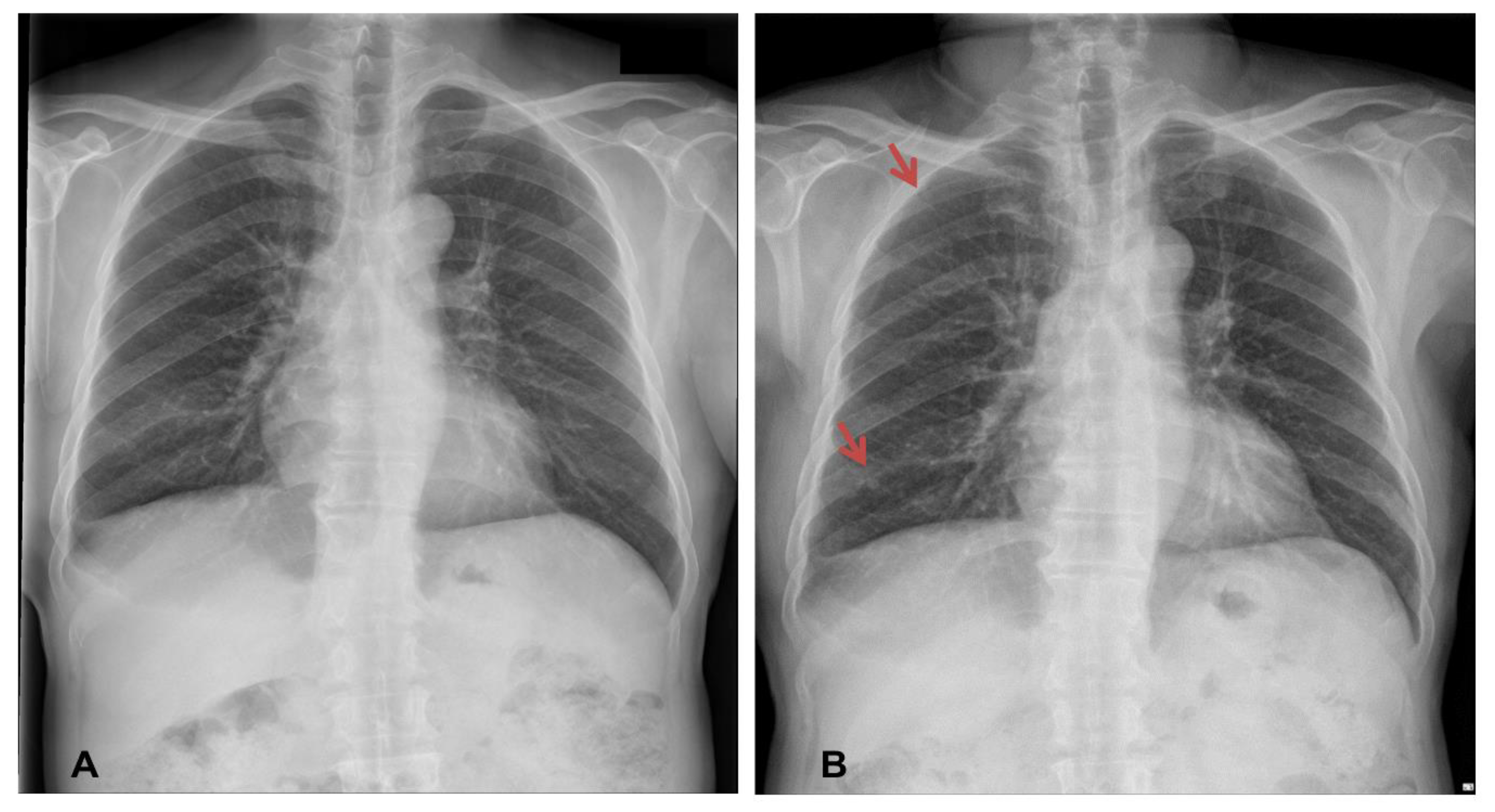
In people who have a history of exposure to asbestos doctors can diagnose an asbestos-related disease with a chest X-ray that shows characteristic changes. A tumor-encased lung appears compressed and can show an elevated diaphragm. Abnormal thickening of the pleura. Renal cell cancer 2. Pleural plaques indicative of asbestos pleural lung disease may also show up in an x-ray. An x-ray may show small irregular opaque areas usually in the lower lobes of the lungs.
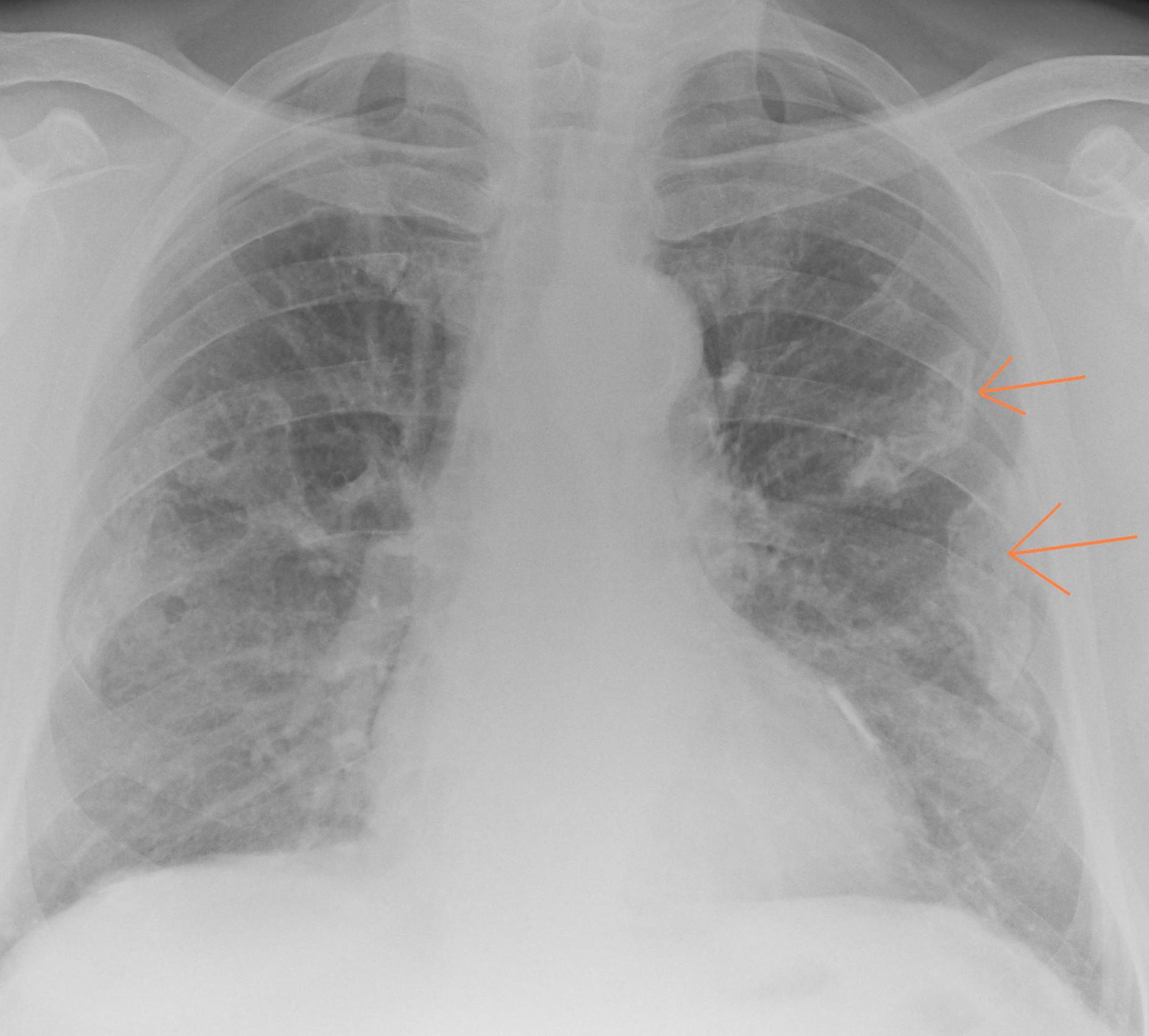
Credit: epos.myesr.org
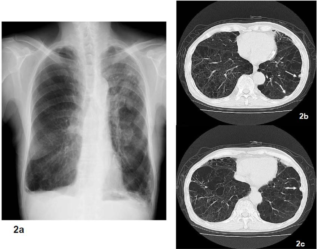
CT however is more sensitive in their detection. CT can help identify the disease in its early stages. Diagnosis of asbestosis is based on history of exposure to asbestos and chest x-ray or high-resolution CT and only rarely requires lung biopsy for confirmation. To be diagnosed with pleural thickening the sufferer will usually attend upon their GP who will take their medical history and carrying out a physical examination. In people who have a history of exposure to asbestos doctors can diagnose an asbestos-related disease with a chest X-ray that shows characteristic changes.

Credit: mdpi.com
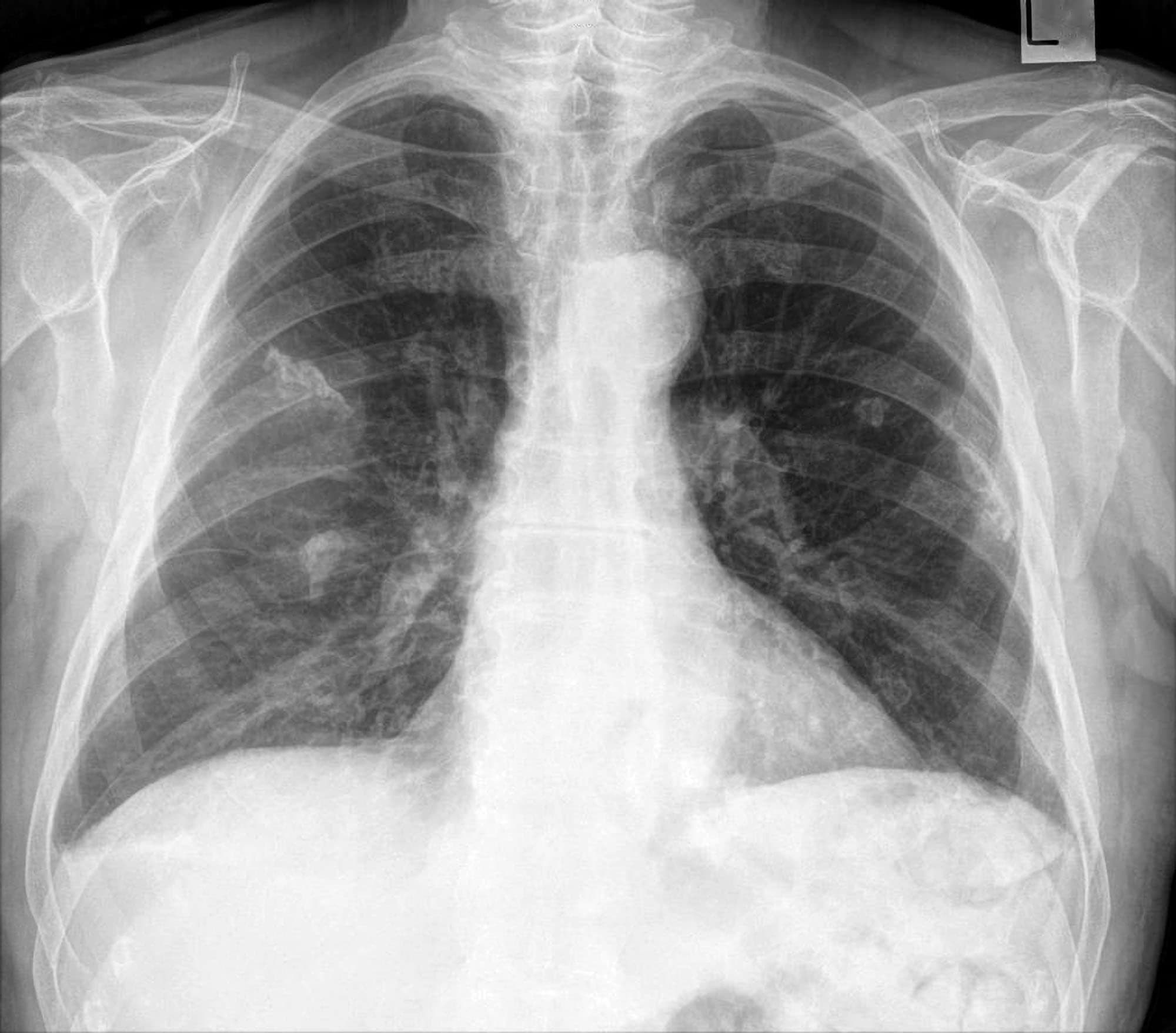
On standard X-rays healthy lungs appear black. When seen en face they may be difficult to see as is the left upper zone plaque in this image. Chest x-ray shows linear reticular opacities signifying fibrosis usually in the peripheral lower lobes. Pleural effusions and pleural plaques are common manifestations of asbestos-related disease. CT can help identify the disease in its early stages.
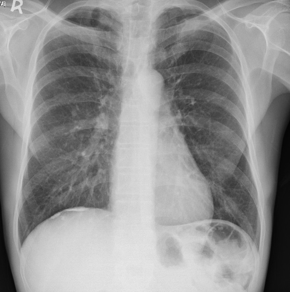
Credit: radiopaedia.org
Diagnosis of asbestosis is based on history of exposure to asbestos and chest x-ray or high-resolution CT and only rarely requires lung biopsy for confirmation. Asbestos related illnesses are usually associated with frequent and. Harron found the X-rays findings were consistent with signs of asbestos-related illness he said he would dictate a report to his staff who would then stamp it with his signature he said. CT can help identify the disease in its early stages. Asbestos-related cancers can occur anywhere in the lungs.

Credit: radiopaedia.org
When seen en face they may be difficult to see as is the left upper zone plaque in this image. An x-ray may show small irregular opaque areas usually in the lower lobes of the lungs. Pleural plaques indicative of asbestos pleural lung disease may also show up in an x-ray. The term pleural asbestosis is not used anymore but rather the term for various types of pleural related and associated scarring is asbestos-related disease including such findings as pleural effusions pleural plaquing diffuse pleural. Abnormal thickening of the pleura.

Credit: medpix.nlm.nih.gov
Pulmonary function was not altered in most of the studied population. The diaphragm is often the best place to look for plaques where they lie in the plane of the X-ray beam. Asbestos plaques - Example 3. Lateral chest xray in asbestosis shows exclusion of alternative plausible causes for the findings. To be diagnosed with pleural thickening the sufferer will usually attend upon their GP who will take their medical history and carrying out a physical examination.
Credit:
In both cases the symptoms can. To be diagnosed with pleural thickening the sufferer will usually attend upon their GP who will take their medical history and carrying out a physical examination. Progressive pulmonary interstitial fibrosis. Diagnosis of asbestosis is based on history of exposure to asbestos and chest x-ray or high-resolution CT and only rarely requires lung biopsy for confirmation. Asbestos plaques - Example 3.
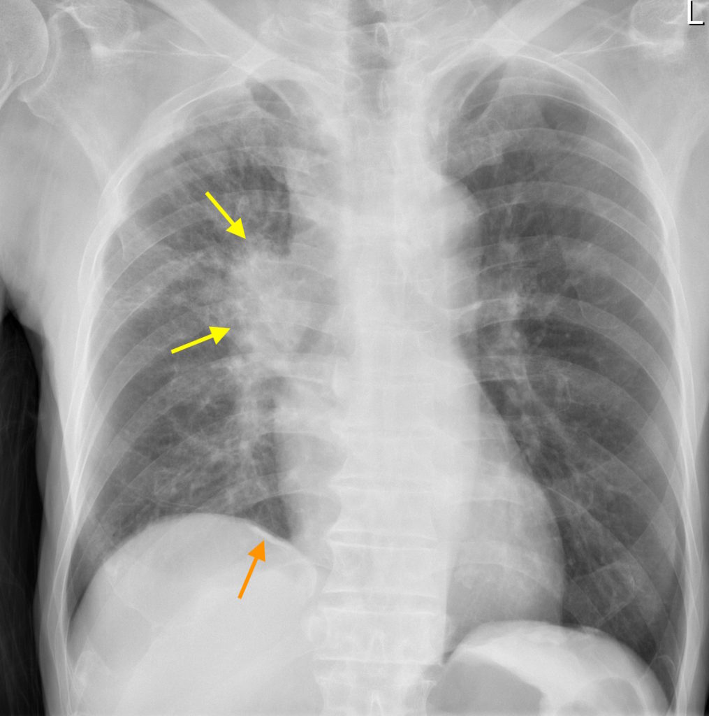
Credit: svuhradiology.ie
Interstitial opacities consistent with fibrosis. The term pleural asbestosis is not used anymore but rather the term for various types of pleural related and associated scarring is asbestos-related disease including such findings as pleural effusions pleural plaquing diffuse pleural. Explore asbestosis x ray findings discover more on when. The numerous incidental findings are a major concern for future screenings which should be considered for asbestos-exposed ex-smokers and current smokers. An exposure to asbestos.
Credit: radiopaedia.org
There are different illnesses one can get from having asbestos in the body not limited to asbestosis and mesothelioma. Diagnosis of asbestosis is based on history of exposure to asbestos and chest x-ray or high-resolution CT and only rarely requires lung biopsy for confirmation. These data are presented in Table III. Case number 7 was the only diagnosed case of asbestosis. CT can help identify the disease in its early stages.
Credit: medschool.co
Pleural effusions and pleural plaques are common manifestations of asbestos-related disease. When seen en face they may be difficult to see as is the left upper zone plaque in this image. When a tumor is present on the pleura doctors will see a wispy white area that indicates tumor growth. Hover onoff image to showhide findings. There are different illnesses one can get from having asbestos in the body not limited to asbestosis and mesothelioma.
Credit: mdpi.com
He was retired for. To be diagnosed with pleural thickening the sufferer will usually attend upon their GP who will take their medical history and carrying out a physical examination. Impairment of the lung function. Lateral chest xray in asbestosis shows exclusion of alternative plausible causes for the findings. Harron found the X-rays findings were consistent with signs of asbestos-related illness he said he would dictate a report to his staff who would then stamp it with his signature he said.
Credit: mdpi.com
It is probable that very shert intervals between first exposure and observed chest x-ray findings indicate causes other than an occupational exposure to asbesos. Asbestos can show up in a chest X-ray as well as other imaging tests for the body according to the Mayo Clinic. Pleural plaques indicative of asbestos pleural lung disease may also show up in an x-ray. An exposure to asbestos. Round atelectasis asbestos pseudotumor.
Credit:
He was retired for. As the fibrosis progresses a number of more definite findings are seen which continue to be particularly subpleural and lower lung zone in distribution. When a tumor is present on the pleura doctors will see a wispy white area that indicates tumor growth. Pleural thickening will usually be diagnosed based on the following findings. Progressive pulmonary interstitial fibrosis.

Credit: onlinelibrary.wiley.com
This was confirmed by chest surgery. Abnormal thickening of the pleura. CT however is more sensitive in their detection. Nineteen of the 148 X-rays had changes consistent with the known prior exposure to asbestos mostly parenchymal in nature. When a tumor is present on the pleura doctors will see a wispy white area that indicates tumor growth.
Credit:
In total 633 workers were included in the present study and were examined with chest radiography and high-resolution CT HRCT. Asbestos plaques - Example 3. As the fibrosis progresses a number of more definite findings are seen which continue to be particularly subpleural and lower lung zone in distribution. In both cases the symptoms can. Interstitial opacities consistent with fibrosis.

Credit: medpix.nlm.nih.gov
Hover onoff image to showhide findings. Impairment of the lung function. Chest radiography in patients with malignant mesothelioma may show an effusion pleural thickening and as the tumor progresses a more lobulated outline. Case number 7 was the only diagnosed case of asbestosis. As the fibrosis progresses a number of more definite findings are seen which continue to be particularly subpleural and lower lung zone in distribution.

Credit: radiopaedia.org
In total 633 workers were included in the present study and were examined with chest radiography and high-resolution CT HRCT. The numerous incidental findings are a major concern for future screenings which should be considered for asbestos-exposed ex-smokers and current smokers. Asbestos can show up in a chest X-ray as well as other imaging tests for the body according to the Mayo Clinic. An x-ray may show small irregular opaque areas usually in the lower lobes of the lungs. Chest x ray findings.

Credit: semanticscholar.org
Renal cell cancer 2. A tumor-encased lung appears compressed and can show an elevated diaphragm. Explore asbestosis x ray findings discover more on when. Radiological findings frequently seen in patients with a history of asbestos exposure include. On standard X-rays healthy lungs appear black.
Credit:
In both cases the symptoms can. The term pleural asbestosis is not used anymore but rather the term for various types of pleural related and associated scarring is asbestos-related disease including such findings as pleural effusions pleural plaquing diffuse pleural. In advanced cases of asbestosis lung tissue may have a honeycomb-like appearance. Interstitial opacities consistent with fibrosis. Asbestosis and first observed x-ray changes.
This site is an open community for users to share their favorite wallpapers on the internet, all images or pictures in this website are for personal wallpaper use only, it is stricly prohibited to use this wallpaper for commercial purposes, if you are the author and find this image is shared without your permission, please kindly raise a DMCA report to Us.
If you find this site adventageous, please support us by sharing this posts to your favorite social media accounts like Facebook, Instagram and so on or you can also bookmark this blog page with the title asbestos x ray findings by using Ctrl + D for devices a laptop with a Windows operating system or Command + D for laptops with an Apple operating system. If you use a smartphone, you can also use the drawer menu of the browser you are using. Whether it’s a Windows, Mac, iOS or Android operating system, you will still be able to bookmark this website.

